")
What is TBI
TBI stands for traumatic brain injury. A traumatic brain injury, more commonly known as a TBI, occurs when an external force comes in contact with the head. The most common TBI injuries are from a car or motorcycle accident, a fall from a roof or ladder, or a jolt in sports.
When there is a direct blow to the head, a bruising of the brain and damage to the internal tissue and blood vessels occurs due to a mechanism called coup-contrecoup. A bruise directly related to trauma at the site of impact is called a coup lesion pronounced, “Coo”. When the brain jolts backwards, it hits the skull on the opposite side and forms a bruise called a contrecoup lesion.

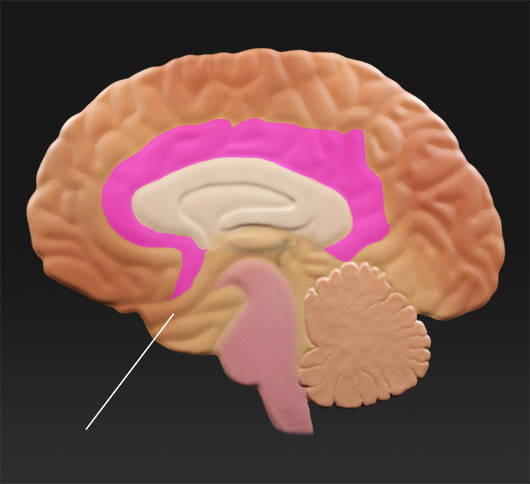
Side View
Frontal Lobe
CerebrumCerebrum
Parietal LobeParietal Lobe
Wernicke's AreaWernicke's Area
Broca's AreaBroca's Area
Olfactory SystemOlfactory System
Temporal LobeTemporal Lobe
CerebellumCerebellum
Occipital LobeOccipital Lobe
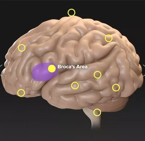
Broca’s Area
Description
Named after 19th-century physician Paul Broca, Broca's area is a section of the brain located in the frontal lobe of the cortex. It is important in the production of written and spoken language.
Function
Broca's area is involved in both written and spoken language processing. It is critical in the complicated task of understandably combining words and grammar. Patients with this condition often do not have a problem with understanding language but have difficulty finding the words or grammar necessary to communicate fluently.
Result of Injury
Injury to this area can cause a condition called Broca's aphasia, also referred to as expressive aphasia, motor aphasia, or non-fluent aphasia. People with this condition are not able to create complex sentences and have difficulty finding the correct word. Patients struggle to form sentences. Their speech is often described as telegraphic, with short sentences containing only keywords. Patients are usually aware that they cannot speak properly.
Example of speech affected by Broca's aphasia: "Ah… Friday… ah, Mom and Steve and Mom… hospital. Two… ah, doctors… and ah… thirty minutes … and yes … ah … hospital. And, er, Wednesday … nine o'clock. And er Thursday, ten o'clock… doctors. Two doctors… and ah… teeth. Yeah, … fine."
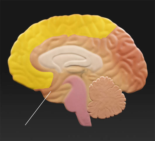
Frontal Lobe
CerebrumCerebrum
Parietal LobeParietal Lobe
Wernicke's AreaWernicke's Area
Broca's AreaBroca's Area
Olfactory SystemOlfactory System
Temporal LobeTemporal Lobe
CerebellumCerebellum
Occipital LobeOccipital Lobe
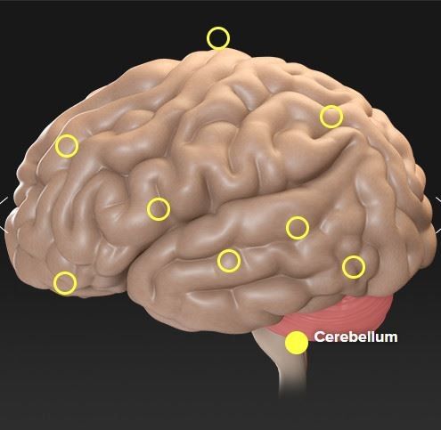
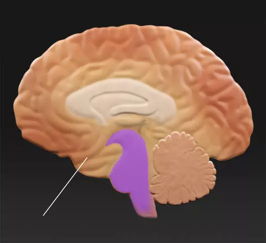
Cerebellum
Description
The word cerebellum means "little brain." The cerebellum is located at the base of the brain and is involved with the coordination of movement. It makes up approximately 10 percent of the brain's volume and contains at least half of the brain's neurons. It is divided into two hemispheres that are covered by a thin layer of gray matter known as the cortex.
Function
The cerebellum receives input from other areas of the brain and spinal cord, along with information about the position and movement of the body's limbs and joints. It uses all of this information to control equilibrium and to provide precise coordination of movements.
Result of Injury
A patient may experience problems with one or more of the following:
Cerebrum
Parietal LobeParietal Lobe
Frontal LobeFrontal Lobe
Wernicke's AreaWernicke's Area
Broca's AreaBroca's Area
Olfactory SystemOlfactory System
Temporal LobeTemporal Lobe
CerebellumCerebellum
Occipital LobeOccipital Lobe
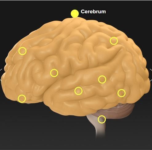
Cerebrum
Description
The cerebrum forms what most people think of as the "brain." It lies on top of the brainstem and is the largest area of the brain. It is divided into two halves — the right hemisphere, which controls the left side of the body, and the left hemisphere, which controls the right side of the body.
Function
The cerebrum's main functions include consciousness, thought, reason, memory, vision, hearing, touch, motor control, coordination, and emotions. The specific functional areas of the cerebrum are divided into four lobes: the frontal, parietal, temporal, and occipital lobes.
Result of Injury
The effect of injury to the cerebrum depends on the specific part of the brain that is injured. In general, injury to the left half of the brain can cause problems on the right side of the body, and damage to the right side of the brain can cause problems on the left side of the body. The results of damage to specific areas of the brain are discussed in the sections for the individual lobes.
Cerebrum
Parietal LobeParietal Lobe
Frontal LobeFrontal Lobe
Wernicke's AreaWernicke's Area
Occipital LobeOccipital Lobe
Broca's AreaBroca's Area
Olfactory SystemOlfactory System
Temporal LobeTemporal Lobe
CerebellumCerebellum
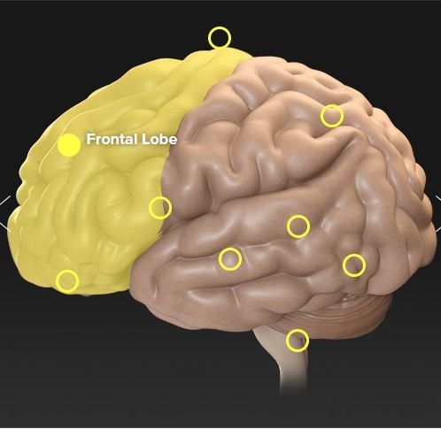
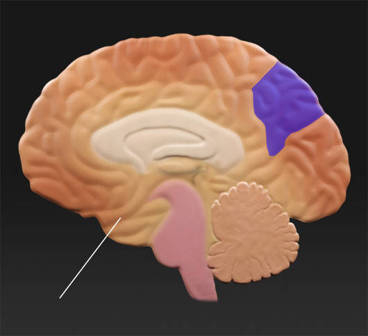
Frontal Lobe
Description
The frontal lobes are located at the front of the cerebrum. They are the part of the brain that regulates and mediates the higher intellectual functions known as executive functions. They play an important role in the control of social behaviors.
Function
The frontal lobes contribute to the regulation of emotions, cognition, error detection, volition, a sense of self, and more. The executive functions of the frontal lobes involve the ability to recognize future consequences resulting from current actions, to choose between good and bad actions, override and suppress unacceptable social responses, and determine similarities and differences between things or events. The frontal lobes also play an important part in retaining longer-term memories, which are often derived from input from the limbic system or emotional brain. The frontal lobe modifies those emotions to generally fit socially acceptable norms.
Result of Injury
Damage to the frontal lobes can lead to a wide variety of symptoms, including the loss of inhibitions and impaired mental flexibility. It may be difficult to prioritize tasks and motivation may diminish. Talking may increase or decrease dramatically. Perceptions about taking risks and following rules can be impaired and socialization can diminish or increase. Damage to some areas of the frontal lobe can affect sexual interest and habits.
Cerebrum
Parietal LobeParietal Lobe
Frontal LobeFrontal Lobe
Wernicke's AreaWernicke's Area
Broca's AreaBroca's Area
Olfactory SystemOlfactory System
Temporal LobeTemporal Lobe
CerebellumCerebellum
Occipital LobeOccipital Lobe
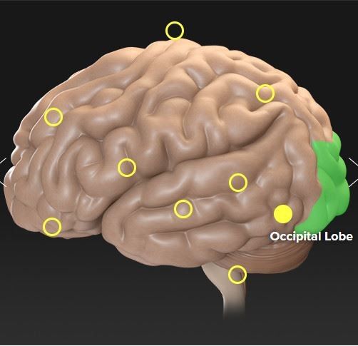
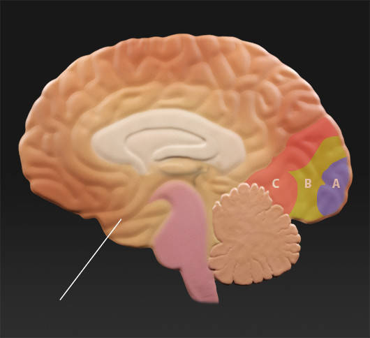
Occipital Lobe
Description
The occipital lobes are located in the back of the brain and are the smallest of the four lobes. They are the visual center and are the primary portion of the brain responsible for receiving input from the eyes.
Function
The occipital lobes receive and process all input from the eyes. Area A is the primary visual cortex and the recipient of visual input from the eyes. Area A performs the initial processing of the information, for example combining input from the two eyes, and beginning to analyze depth. This information is then passed on to Areas B and C. Areas B and C are the associative visual cortex, where motion, color, and other parameters are interpreted to provide meaning.
Result of the Injury
Damage to the right occipital lobe can result in loss of the left visual field of both eyes, and damage to the left occipital lobe results in loss of the right visual field of both eyes. Damage to both the left and right occipital lobes can result in blindness, even though the eyes themselves may be functioning normally. This condition is called cortical blindness. If the front part of the occipital lobe is damaged, people have difficulty recognizing familiar objects and faces and accurately interpreting what they see.
Cerebrum
Parietal LobeParietal Lobe
Frontal LobeFrontal Lobe
Wernicke's AreaWernicke's Area
Broca's AreaBroca's Area
Olfactory SystemOlfactory System
Temporal LobeTemporal Lobe
CerebellumCerebellum
Occipital LobeOccipital Lobe
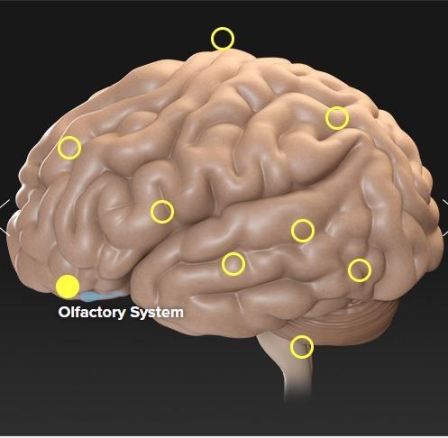
Olfactory System
Description
The olfactory system includes the olfactory epithelium, which is located inside the nose, the olfactory bulb, which is located just above the olfactory epithelium, and the olfactory cortex, which is on the surface of the temporal lobe. The olfactory system is responsible for the sensing, processing, and identifying of smells.
Function
The olfactory system is responsible for the sense of smell. In addition, it combines with taste information in the brain to create the sensation of flavor. Many people with a lost sense of smell retain the ability to detect salty, sweet, and bitter tastes but complain that food tastes bland. The sense of smell is strongly associated with memory. As a result, smells are often powerful triggers for specific memories, both good and bad. Because the sense of smell is also closely associated with the limbic system or emotional brain, the smell is known to provoke strong emotional responses.
Result of Injury
Damage to the olfactory system can cause impairment or loss of the sense of smell and the sense of taste. Much of what we attribute to our sense of taste actually depends on the aromas of what we eat and drink.
Cerebrum
Parietal LobeParietal Lobe
Frontal LobeFrontal Lobe
Wernicke's AreaWernicke's Area
Broca's AreaBroca's Area
Olfactory SystemOlfactory System
Temporal LobeTemporal Lobe
CerebellumCerebellum
Occipital LobeOccipital Lobe
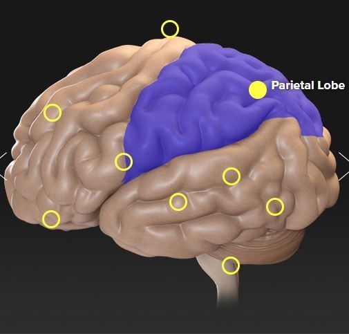
Parietal Lobe
Description
The parietal lobes are positioned above the temporal lobes and between the frontal and occipital lobes. They contain the part of the brain primarily responsible for movement and the sense of touch. They also play a very important role in integrating sensory information, particularly determining spatial sense and navigation.
Function
The parietal lobes contain the portion of the brain primarily responsible for movement. This area is called the motor cortex. Located next to the motor cortex is the area of the brain primarily responsible for the sense of touch. This area is known as the somatosensory cortex. A large portion of the parietal lobe acts as an interchange for information for most of the other senses. These interconnections contribute a great deal to the ability to interpret people, places, and things around us. In essence, the parietal lobes "pull it all together."
Result of Injury
Damage to the parietal lobes can result in conditions known as agnosia and apraxia. Agnosia is the inability to recognize an object using a specific sense, even though that sense is basically intact. For example, someone might not be able to recognize an orange by sight, although they could identify the orange by touch or smell.
Apraxia is the inability to perform a specific movement, although all of the muscles are intact and able to perform that task under different circumstances. For example, someone may be unable to touch their nose on command but would be perfectly able to do so if their nose itched.
Damage to the parietal lobe can also cause word blindness, known as alexia, with writing impairments, called agraphia. Damage to the right parietal lobe can impair skills such as dressing and washing, and can also cause difficulty in making things, denial of deficits, and loss of drawing ability. Damage can also cause a person to neglect the opposite side of their body, even to the point of not recognizing their own limbs.
Cerebrum
Parietal LobeParietal Lobe
Frontal LobeFrontal Lobe
Wernicke's AreaWernicke's Area
Broca's AreaBroca's Area
Olfactory SystemOlfactory System
Temporal LobeTemporal Lobe
CerebellumCerebellum
Occipital LobeOccipital Lobe
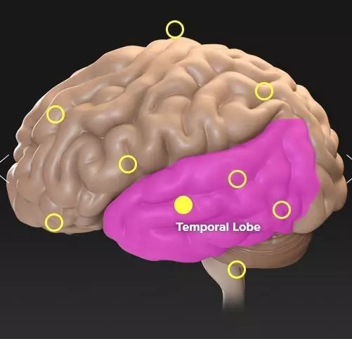
Temporal Lobe
Description
The temporal lobes are the part of the cerebrum that is involved in speech, memory, and hearing. They are located on both sides of the brain, below the parietal lobes and in front of the occipital lobes.
Function
The temporal lobe is involved in hearing and understanding what is heard and is home to the primary auditory cortex. It is also responsible for the formation and retrieval of long-term memories. The temporal lobe also plays a role in vision, sexual behavior, and personality.
Result of Injury
Individuals with temporal lobe damage have difficulty placing words or pictures into categories. Language skills can also be impaired by temporal lobe damage. The impairment or loss of long-term memory can result from injury to the temporal lobes. Specifically, damage to the right side of the temporal lobes can result in the inability to recall non-verbal material, such as music and drawings. It can also cause a loss of inhibition when talking. Left temporal lobe damage can impair the ability to recognize words and can result in impaired memory for verbal material.
Cerebrum
Parietal LobeParietal Lobe
Frontal LobeFrontal Lobe
Wernicke's AreaWernicke's Area
Broca's AreaBroca's Area
Olfactory SystemOlfactory System
Temporal LobeTemporal Lobe
CerebellumCerebellum
Occipital LobeOccipital Lobe
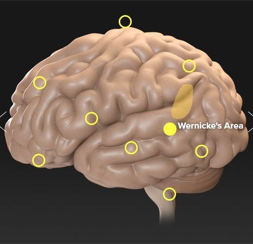
Wernicke’s Area
Description
Wernicke's area is located in the left temporal lobe. It plays a role in the understanding of language and is named after Carl Wernicke, a German neurologist, and psychiatrist. In 1874, Wernicke discovered that damage to this area could cause a type of speech disorder that is now called Wernicke's aphasia.
Function
Wernicke's area plays an important role in the processing and comprehension of language. Proper function of Wernicke's area is required for patients to form coherent sentences and to understand the speech of others.
Result of Injury
Damage to Wernicke's area can cause a condition called Wernicke's aphasia or receptive aphasia. This condition affects a person's ability to understand the language when listening, the ability to read, and the ability to speak with meaning. Speech often sounds like a babble, fluent and grammatical, though totally nonsensical. Wernicke's aphasics have severe difficulties with comprehension but are often unaware of it. They may become annoyed or frustrated when others can't seem to understand them.
Example of speech affected by Wernicke's aphasia: "I can't tell you what that is, but I know what it is, but I don't know where it is. But I don't know what's under. I know it's you couldn't say it's… I couldn't say what it is. I couldn't say what that is. This shu… that should be right in here. That's very bad in there. Anyway, this one here, and that, and that's it. This is the getting in here and that's the getting around here, and that, and that's it. This is getting in here and that's the getting around here, this one and one with this one. And this one, and that's it, isn't it? I don't know what else you'd want."
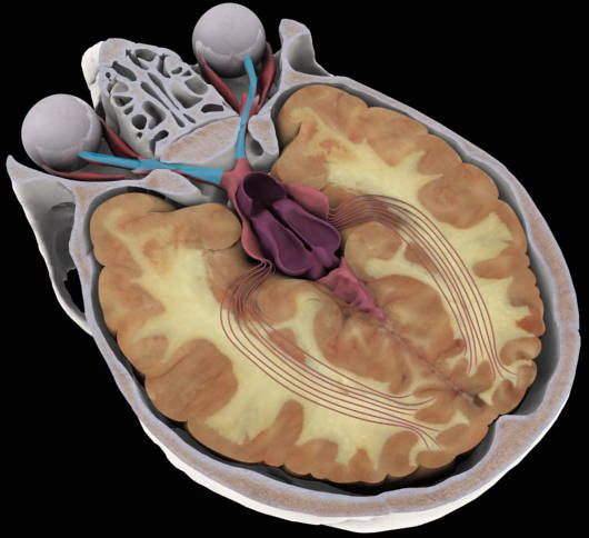
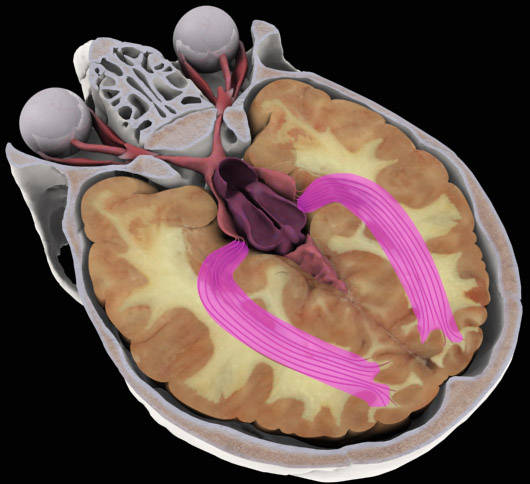
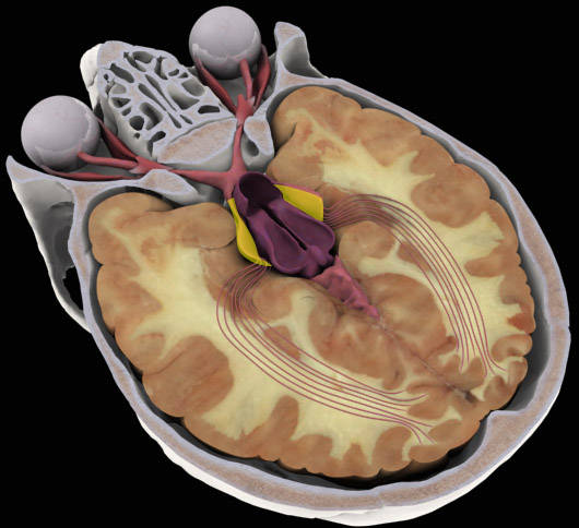
Inside View
Brain Stem
CerebellumCerebellum
Frontal LobeFrontal Lobe
HypothalamusHypothalamus
Limbic SystemLimbic System
Occipital LobeOccipital Lobe
Olfactory SystemOlfactory System
Parietal LobeParietal Lobe
Septal (Corpus Collosum)Septal (Corpus Collosum)
Temporal LobeTemporal Lobe
Brain Stem
Description
The brain stem is a part of the brain located beneath the cerebrum and in front of the cerebellum. It connects the spinal cord to the rest of the brain and is responsible for regulating many of the body's basic functions. It is sometimes referred to as the most primitive portion of the brain.
Function
Neurological functions located in the brainstem include those necessary for survival such as breathing, digestion, heart rate, and blood pressure, and for being awake and alert. It plays an important role in basic attention, arousal, and consciousness. One key part of the brain stem is called the reticular activating system, which works as the "on/off switch," in terms of sleeping, waking up, and motivation.
The brain stem serves as a conduit, with information to and from other parts of the body passing through the brain stem on the way to or from the brain.
Many of the cranial nerves originate in or pass through the brain stem. These nerves are responsible for things such as vision, hearing, taste, smell, the control of eye movements, and movement and sensation of the face.
Result of Injury
The brain stem is vital to proper body functions and can cause severe disabilities if it is damaged. In fact, because the brain stem controls such basic functions as breathing and heartbeat, damage to the brain stem can result in death. Brain stem injury is also a cause for persistent coma. Brain stem injury can also result in memory problems, difficulty concentrating, difficulty staying focused, and physical problems including the inability to walk, remain balanced, and a loss of strength. Problems with vision, such as impaired eye movement, can also be caused by injury to the brain stem. Paralysis may also occur when the brain stem is damaged since it is directly linked to the spinal cord.
Brain Stem
CerebellumCerebellum
Frontal LobeFrontal Lobe
HypothalamusHypothalamus
Limbic SystemLimbic System
Occipital LobeOccipital Lobe
Olfactory SystemOlfactory System
Parietal LobeParietal Lobe
Septal (Corpus Collosum)Septal (Corpus Collosum)
Temporal LobeTemporal Lobe
Cerebellum
Description
The word cerebellum means "little brain." The cerebellum is located at the base of the brain and is involved with the coordination of movement. It makes up approximately 10 percent of the brain's volume and contains at least half of the brain's neurons. It is divided into two hemispheres that are covered by a thin layer of gray matter known as the cortex.
Function
The cerebellum receives input from other areas of the brain and spinal cord, along with information about the position and movement of the body's limbs and joints. It uses all of this information to control equilibrium and to provide precise coordination of movements.
Result of Injury
A patient may experience problems with one or more of the following:
Brain Stem
CerebellumCerebellum
Frontal LobeFrontal Lobe
HypothalamusHypothalamus
Limbic SystemLimbic System
Occipital LobeOccipital Lobe
Olfactory SystemOlfactory System
Parietal LobeParietal Lobe
Septal (Corpus Collosum)Septal (Corpus Collosum)
Temporal LobeTemporal Lobe
Frontal Lobe
Description
The frontal lobes are located at the front of the cerebrum. They are the part of the brain that regulates and mediates the higher intellectual functions known as executive functions. They play an important role in the control of social behaviors.
Function
The frontal lobes contribute to the regulation of emotions, cognition, error detection, volition, a sense of self, and more. The executive functions of the frontal lobes involve the ability to recognize future consequences resulting from current actions, to choose between good and bad actions, override and suppress unacceptable social responses, and determine similarities and differences between things or events. The frontal lobes also play an important part in retaining longer-term memories, which are often derived from input from the limbic system or emotional brain. The frontal lobe modifies those emotions to generally fit socially acceptable norms.
Result of Injury
Damage to the frontal lobes can lead to a wide variety of symptoms, including the loss of inhibitions and impaired mental flexibility. It may be difficult to prioritize tasks and motivation may diminish. Talking may increase or decrease dramatically. Perceptions about taking risks and following rules can be impaired and socialization can diminish or increase. Damage to some areas of the frontal lobe can affect sexual interest and habits.
Brain Stem
CerebellumCerebellum
Frontal LobeFrontal Lobe
HypothalamusHypothalamus
Limbic SystemLimbic System
Occipital LobeOccipital Lobe
Olfactory SystemOlfactory System
Parietal LobeParietal Lobe
Septal (Corpus Collosum)Septal (Corpus Collosum)
Temporal LobeTemporal Lobe
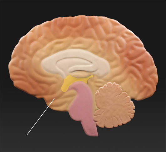
Hypothalamus
Description
The hypothalamus links the nervous system to the endocrine system, which is primarily responsible for metabolism. It is located just above the brain stem.
Function
The main function of the hypothalamus is to maintain the body's status quo. The hypothalamus precisely controls blood pressure, body temperature, fluid and electrolyte levels, and body weight. It also influences hunger, moods, sex drive, circadian cycles, thirst, and the release of hormones from the pituitary gland.
Result of Injury
Injury to the hypothalamus can result in a wide range of symptoms, including weight loss or gain, depression, aggression, insomnia, loss of sex drive, hypersexuality, and many other conditions. Severe injury to the hypothalamus, such as a shearing injury, can be fatal because it can disrupt the normal homeostasis of blood chemistry, body temperature, and other important functions.
Brain Stem
CerebellumCerebellum
Frontal LobeFrontal Lobe
HypothalamusHypothalamus
Limbic SystemLimbic System
Occipital LobeOccipital Lobe
Olfactory SystemOlfactory System
Parietal LobeParietal Lobe
Septal (Corpus Collosum)Septal (Corpus Collosum)
Temporal LobeTemporal Lobe
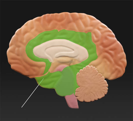
Limbic System
Description
The limbic system lies just under the cerebrum within the core of the brain. It is thought to include the hypothalamus, the hippocampus, and the amygdala, and other structures. The role of the limbic system is not well understood; however, it plays an important part in regulating emotions and pleasure and contributes to the formation of memories.
Function
The limbic system is involved in emotions and motivation, especially those related to survival, including fear and anger. The limbic system is also involved in feelings of pleasure, such as those experienced from eating and sexual activity. The amygdala and hippocampus are key parts of the limbic system that play important roles in memory. The amygdala is responsible for determining what memories are stored and wherein the brain those memories are stored.
Result of Injury
Injury to the limbic system can result in changes in behaviors ranging from subtle to severe. Those changes can include such things as flat affect, which means showing no expression, aggressiveness, mild distractibility, or the inability to pay attention for any length of time. Memory problems can also result from injury to the limbic system, ranging from almost imperceptible challenges with the recall to the inability to remember an event that just happened.
Brain Stem
CerebellumCerebellum
Frontal LobeFrontal Lobe
HypothalamusHypothalamus
Limbic SystemLimbic System
Occipital LobeOccipital Lobe
Olfactory SystemOlfactory System
Parietal LobeParietal Lobe
Septal (Corpus Collosum)Septal (Corpus Collosum)
Temporal LobeTemporal Lobe
Occipital Lobe
Description
The occipital lobes are located in the back of the brain and are the smallest of the four lobes. They are the visual center and are the primary portion of the brain responsible for receiving input from the eyes.
Function
The occipital lobes receive and process all input from the eyes. Area A is the primary visual cortex and the recipient of visual input from the eyes. Area A performs the initial processing of the information, for example combining input from the two eyes, and beginning to analyze depth. This information is then passed on to Areas B and C. Areas B and C are the associative visual cortex, where motion, color, and other parameters are interpreted to provide meaning.
Result of the Injury
Damage to the right occipital lobe can result in loss of the left visual field of both eyes, and damage to the left occipital lobe results in loss of the right visual field of both eyes. Damage to both the left and right occipital lobes can result in blindness, even though the eyes themselves may be functioning normally. This condition is called cortical blindness. If the front part of the occipital lobe is damaged, people have difficulty recognizing familiar objects and faces and accurately interpreting what they see.

Brain Stem
CerebellumCerebellum
Frontal LobeFrontal Lobe
HypothalamusHypothalamus
Limbic SystemLimbic System
Occipital LobeOccipital Lobe
Olfactory SystemOlfactory System
Parietal LobeParietal Lobe
Septal (Corpus Collosum)Septal (Corpus Collosum)
Temporal LobeTemporal Lobe
Olfactory System
Description
The olfactory system includes the olfactory epithelium, which is located inside the nose, the olfactory bulb, which is located just above the olfactory epithelium, and the olfactory cortex, which is on the surface of the temporal lobe. The olfactory system is responsible for the sensing, processing, and identifying of smells.
Function
The olfactory system is responsible for the sense of smell. In addition, it combines with taste information in the brain to create the sensation of flavor. Many people with a lost sense of smell retain the ability to detect salty, sweet, and bitter tastes, but complain that food tastes bland. The sense of smell is strongly associated with memory. As a result, smells are often powerful triggers for specific memories, both good and bad. Because the sense of smell is also closely associated with the limbic system or emotional brain, the smell is known to provoke strong emotional responses.
Result of Injury
Damage to the olfactory system can cause impairment or loss of the sense of smell and the sense of taste. Much of what we attribute to our sense of taste actually depends on the aromas of what we eat and drink.
Brain Stem
CerebellumCerebellum
Frontal LobeFrontal Lobe
HypothalamusHypothalamus
Limbic SystemLimbic System
Occipital LobeOccipital Lobe
Olfactory SystemOlfactory System
Parietal LobeParietal Lobe
Septal (Corpus Collosum)Septal (Corpus Collosum)
Temporal LobeTemporal Lobe
Parietal Lobe
Description
The parietal lobes are positioned above the temporal lobes and between the frontal and occipital lobes. They contain the part of the brain primarily responsible for movement and the sense of touch. They also play a very important role in integrating sensory information, particularly determining spatial sense and navigation.
Function
The parietal lobes contain the portion of the brain primarily responsible for movement. This area is called the motor cortex. Located next to the motor cortex is the area of the brain primarily responsible for the sense of touch. This area is known as the somatosensory cortex. A large portion of the parietal lobe acts as an interchange for information for most of the other senses. These interconnections contribute a great deal to the ability to interpret people, places, and things around us. In essence, the parietal lobes "pull it all together."
Result of Injury
Damage to the parietal lobes can result in conditions known as agnosia and apraxia. Agnosia is the inability to recognize an object using a specific sense, even though that sense is basically intact. For example, someone might not be able to recognize an orange by sight, although they could identify the orange by touch or smell.
Apraxia is the inability to perform a specific movement, although all of the muscles are intact and able to perform that task under different circumstances. For example, someone may be unable to touch their nose on command but would be perfectly able to do so if their nose itched.
Damage to the parietal lobe can also cause word blindness, known as alexia, with writing impairments, called agraphia. Damage to the right parietal lobe can impair skills such as dressing and washing, and can also cause difficulty in making things, denial of deficits, and loss of drawing ability. Damage can also cause a person to neglect the opposite side of their body, even to the point of not recognizing their own limbs.
Brain Stem
CerebellumCerebellum
Frontal LobeFrontal Lobe
HypothalamusHypothalamus
Limbic SystemLimbic System
Occipital LobeOccipital Lobe
Olfactory SystemOlfactory System
Parietal LobeParietal Lobe
Septal (Corpus Collosum)Septal (Corpus Collosum)
Temporal LobeTemporal Lobe
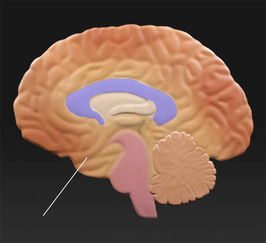
Septal (Corpus Collosum)
Description
The corpus callosum is a thick band of fibers located between the cerebral hemispheres. It connects the left and right sides of the brain and is the largest fiber bundle in the human brain, with more than 300 million axons.
Function
The job of the corpus callosum is to channel nerve transmissions between the two hemispheres of the brain. It ensures that each hemisphere has access to data from the opposite side of the body and the surrounding environment.
Result of Injury
Damage to the front of the corpus callosum can lead to impaired judgment and defective memory, while damage to the rear part can cause behavioral changes.
Brain Stem
CerebellumCerebellum
Frontal LobeFrontal Lobe
HypothalamusHypothalamus
Limbic SystemLimbic System
Occipital LobeOccipital Lobe
Olfactory SystemOlfactory System
Parietal LobeParietal Lobe
Septal (Corpus Collosum)Septal (Corpus Collosum)
Temporal LobeTemporal Lobe
Temporal Lobe
Description
The temporal lobes are the part of the cerebrum that is involved in speech, memory, and hearing. They are located on both sides of the brain, below the parietal lobes and in front of the occipital lobes.
Function
The temporal lobe is involved in hearing and understanding what is heard and is home to the primary auditory cortex. It is also responsible for the formation and retrieval of long-term memories. The temporal lobe also plays a role in vision, sexual behavior, and personality.
Result of Injury
Individuals with temporal lobe damage have difficulty placing words or pictures into categories. Language skills can also be impaired by temporal lobe damage. The impairment or loss of long-term memory can result from injury to the temporal lobes. Specifically, damage to the right side of the temporal lobes can result in the inability to recall non-verbal material, such as music and drawings. It can also cause a loss of inhibition when talking. Left temporal lobe damage can impair the ability to recognize words and can result in impaired memory for verbal material.
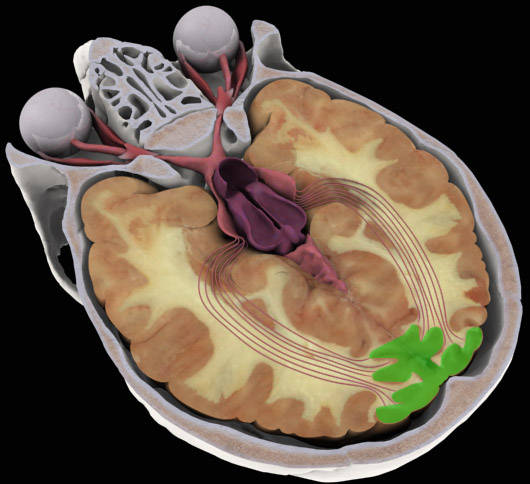
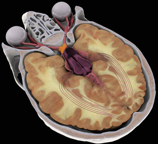
Visual Anatomy
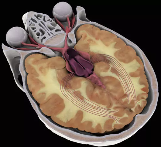
Eyes
Occipital LobeOccipital Lobe
Optic ChiasmOptic Chiasm
Optic NervesOptic Nerves
Optic RadiationsOptic Radiations
Optic TractOptic Tract
Eyes
Description
The eye is the body's link to the visible environment. It is a complex organ designed to collect and focus light, which is then converted by the retina into an electrical signal and sent to the occipital lobe of the brain for processing and interpretation.
Eyes
Occipital LobeOccipital Lobe
Optic ChiasmOptic Chiasm
Optic NervesOptic Nerves
Optic RadiationsOptic Radiations
Optic TractOptic Tract
Occipital Lobe
Description
The occipital lobes are located in the back of the brain and are the smallest of the four lobes. They are the visual center and are the primary portion of the brain responsible for receiving input from the eyes.
Function
The occipital lobes receive and process all input from the eyes. Area A is the primary visual cortex and the recipient of visual input from the eyes. Area A performs the initial processing of the information, for example combining input from the two eyes, and beginning to analyze depth. This information is then passed on to Areas B and C. Areas B and C are the associative visual cortex, where motion, color, and other parameters are interpreted to provide meaning.
Result of the Injury
Damage to the right occipital lobe can result in loss of the left visual field of both eyes, and damage to the left occipital lobe results in loss of the right visual field of both eyes. Damage to both the left and right occipital lobes can result in blindness, even though the eyes themselves may be functioning normally. This condition is called cortical blindness. If the front part of the occipital lobe is damaged, people have difficulty recognizing familiar objects and faces and accurately interpreting what they see.
Eyes
Occipital LobeOccipital Lobe
Optic ChiasmOptic Chiasm
Optic NervesOptic Nerves
Optic RadiationsOptic Radiations
Optic TractOptic Tract
Optic Chiasm
Description
The optic chiasm is the point where the two optic nerves intersect. In the chiasm, part of the nerve fibers from each optic nerve cross to the opposite side of the brain and combine with the remaining fibers from the other optic nerve. This results in most of the right visual field from both eyes going to the left side of the brain and most of the left visual field from both eyes going to the right side of the brain. At this point, injuries change from affecting the visual field of one eye to affecting both eyes.
Eyes
Occipital LobeOccipital Lobe
Optic ChiasmOptic Chiasm
Optic NervesOptic Nerves
Optic RadiationsOptic Radiations
Optic TractOptic Tract
Optic Nerves
Description
The optic nerve is the large nerve bundle that exits from the back of the eye. It is made up of over 2 million individual nerves, measures about 4mm in diameter, and carries the electrical signals from the eye to the brain. Head trauma can damage the optic nerve in several ways. A blow to the head can disrupt the blood flow to the optic nerve or it can cause a bone to break and press on the optic nerve. This type of injury is usually called traumatic optic neuropathy and can result in partial to complete loss of vision in the affected eye.
Eyes
Occipital LobeOccipital Lobe
Optic ChiasmOptic Chiasm
Optic NervesOptic Nerves
Optic RadiationsOptic Radiations
Optic TractOptic Tract
OPTIC RADIATIONS
Description
The part of the visual pathway that extends from the lateral geniculate nuclei to the occipital lobes is called optic radiations. The optic radiations carry the electrical signals that originated in the eye at the final distance to the occipital lobes.
Eyes
Occipital LobeOccipital Lobe
Optic ChiasmOptic Chiasm
Optic NervesOptic Nerves
Optic RadiationsOptic Radiations
Optic TractOptic Tract
Optic Tract
Description
The combined nerve fibers from the two optic nerves form the optic tract on each side of the brain. The optic tracts extend from the optic chiasm to a point midway to the occipital lobe of the brain called the lateral geniculate nucleus.
Motility

Normal Eye Movement
Description
With normal eye movements, the eyes are aligned to focus on the same object and provide a single image to the brain. Both eyes track together when looking in any direction. Each eye has six muscles and three nerves that control its movements. Misalignment of the eyes can result in double vision. Regardless of the suspected cause, all cases of double vision must be evaluated by a doctor.
Function
Eye movements are very complex and carefully controlled by the brain. Rapid movements, called saccades, are required to quickly change fixation from one object to another. Slower, smooth eye movements called pursuits are required to track moving objects or follow words on a page to read. When looking at an object up-close, both eyes turn toward the nose. This movement is called convergence.
Result of Injury
Proper coordination of eye movements can be disturbed in a number of ways. TBI alone can result in a decreased ability of the brain to control fine movements, such as saccades and pursuits. This type of injury can interfere with reading or activities involving fast-moving objects. Damage to one of the eye muscles or to the nerve that controls the muscle can lead to misalignment of the eyes. This type of injury results in seeing two objects when only one exists and is called double vision or diplopia. Some injuries improve overtime or the effects can be lessened with surgery on the eye muscles. Other injuries are permanent and ways to lessen their impact must be developed with the healthcare team.
Cranial Nerve III Injury (Oculomotor)
Description
Cranial Nerve III is also known as the oculomotor nerve. This nerve controls four of the six muscles attached to each eye. It also controls the elevation of the eyelid and constriction of the pupil.
Function
The oculomotor nerve controls the superior rectus muscle (up gaze), medial rectus muscle (inward gaze), inferior rectus muscle (downgaze), inferior oblique muscle (up and inward gaze, rotation of the eye), and the levator palpebrae superioris (raises upper eyelid). Special nerve fibers associated with cranial nerve III control constriction of the pupil.
Result of Injury
Traumatic injury to the cranial nerve III results in the eye remaining in a down and out position, the upper eyelid is droopy and the pupil may be large. Double vision is present if the eyelid is lifted. Due to the large number of muscles affected, a surgical correction is rarely an option. People with diabetes can experience a temporary loss of function of this nerve but with a normal pupil. This condition usually resolves over a period of time.
Cranial Nerve IV Injury (Trochlear)
Description
Cranial Nerve IV is also known as the trochlear nerve. This nerve controls the superior oblique muscle. The name trochlear nerve comes from the small pulley-like structure the tendon of the superior oblique muscle passes through called the trochlea. Most cranial nerve IV palsies are congenital.
Function
The trochlear nerve controls the superior oblique muscle. This muscle is responsible for the down and inward movement of the eye as well as rotation of the eye when the head is tilted.
Result of Injury
A traumatic injury to cranial nerve IV will cause the affected eye to be in more of an up gaze compared to the normal eye. This is worsened when attempting to look down and in with the injured eye. Double vision is common and the images are often diagonally spaced. To reduce double vision, people with an injury to the trochlear nerve will often tilt their head toward the shoulder opposite from the injury.
Cranial Nerve VI Injury (Abducens)
Description
Cranial Nerve VI is also known as the abducens nerve. This nerve controls the lateral rectus muscle and is responsible for lateral or outward movement of the eye. This movement is known as abduction and is the source for the name of the nerve.
Function
The abducens nerve controls the lateral rectus muscle, which controls the lateral or outward movement of the eye. In other words, it controls the left eye's left gaze and the right eye's right gaze.
Result of Injury
A traumatic injury to cranial nerve VI will cause the affected eye to be turned in toward the nose when compared to the normal eye. When attempting to look laterally, the affected eye will not go past its center position. Double vision is common, and the images are often horizontally spaced. To reduce double vision, people with an injury to the abducens nerve will often turn their heads toward the side with the nerve injury.
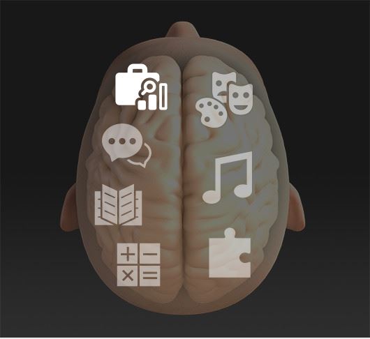
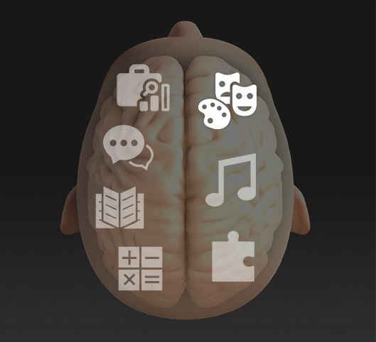
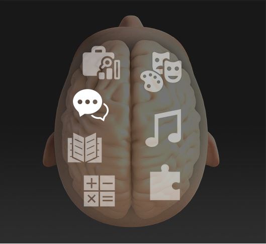
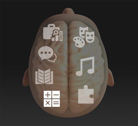
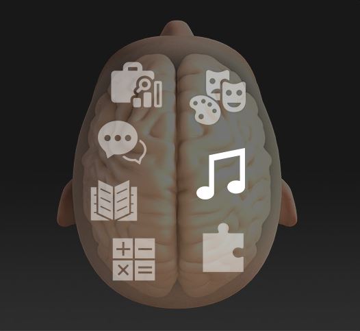
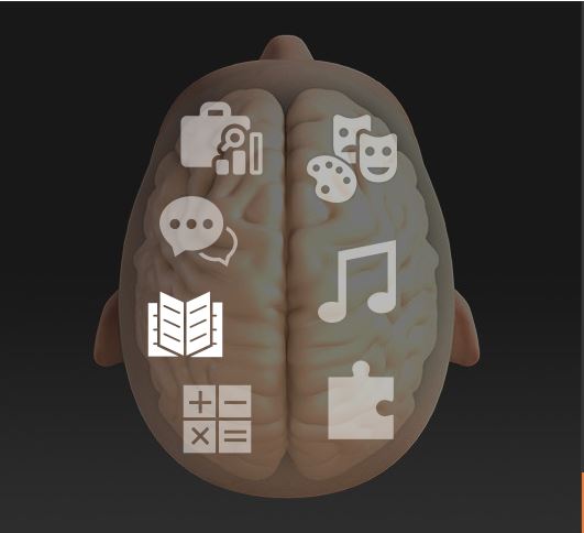
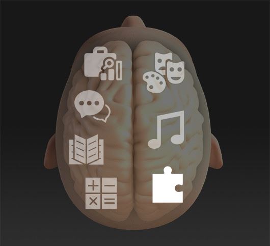
Left and Right Brain Function
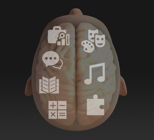
Left and Right Brain
Description
LEFT BRAIN: In most people, the left hemisphere of the brain is dominant for language, mathematical skills, and the ability to solve problems in sequential order. For this reason, the left hemisphere is often considered the logical or analytical brain.
RIGHT BRAIN: The right hemisphere of the brain seems to be dominant in terms of artistic ability, musical skills, face recognition, and spatial perception. Problems tend to be solved in a more comprehensive, holistic manner by the right brain.